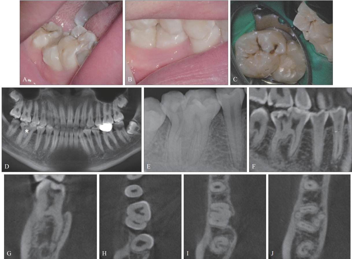
右下颌融合磨牙7根管显微根管治疗1例

打开文本图片集
[中图分类号] R781.05 [文献标志码] B [doi] 10.7518/hxkq.2025.2024408
[Abstract]Fused teeth are usually formed bythe partial orcomplete fusionof two normal tooth germs during the developmentprocessandbelong todentaldevelopmentalabnormalities.Fusedteetharerelativelyrareclinically,ndthose occurring in the posterior tooth area areevenrarer.This articlereports acase offused teth between the firstpermanent molarand the second permanent molar in the right mandible.This fusedtooth hadacomplex rootcanal anatomical structure (seven root canals).The number and locationof the root canals were analyzed by cone beam computed tomography, and root canal treatment was successfully completed with the assistance of microscope.
[Keywords] fused teeth; canal anatomy;cone beam computed tomography;microscopic root canal treatment
牙齿的发育异常可表现为大小、形状、数量或结构等多个方面。(剩余8390字)
