心电瀑布图诊断间歌性QRS波增宽的临床价值

打开文本图片集
[中图分类号] R540.41[文献标志码]A [文章编号] 2097-5716(2025)03-0409-09 DOI: 10.13308/j.issn.2097-5716.2025.03.019
[引用格式]王晶晶,景永明,申继红,等.心电瀑布图诊断间歇性QRS 波增宽的临床价值[J].实用心电与临床诊疗,2025,34(3):409-417.
Clinical value of electrocardiographic waterfall plots in diagnosing intermitent QRS complex widening
WANG Jingjing’, JING Yongming’, SHEN Jihong',LIU Shichao', GENG Yiming²,LI Shifeng1(Department of Electrocardiography,1.the Second Affiliated Hospital of Zhengzhou University, Zhengzhou Henan 450O14;2.the Fifth Affiliated Hospital of Zhengzhou University, Zhengzhou Henan 45OO15,China)
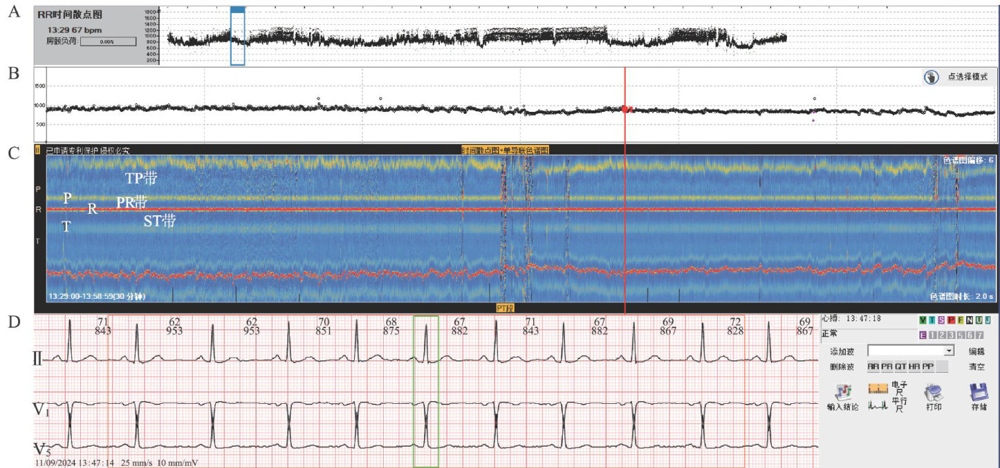
[Abstract]ObjectiveTo explore the clinical value of electrocardiographic waterfall plots in the diagnosis of intermittent QRS complex widening. MethodsThe typical cases with intermitent QRS complex widening in ambulatory electrocardiography were retrospectively analyzed.The characteristics of their electrocardiographic waterfall plots were observed and summarized.ResultsDuring intermitent QRS complex widening,the electrocardiographic waterfall plot demonstrated wideningand discoloration of the R-peak band with concomitant T-peak band discoloration. The electrocardiographic patern showing normal P-peak band progression with a PR interval within anormal range wasobserved in conditions suchas intermitent left orright bundle branch block and VAT ventricular pacing.Abnormal P-peak band progresion (manifesting as shift,fragmentation,or absence)was observed inidioventricular rhythms and VVI pacing rhythms;whereas normal P-peak band progression with shortenedPR intervalsoccurred in intermittentventricular prexcitation.ConclusionElectrocardiographic waterfall plotissuitableforrapid diagnosisanddiffrentialdiagnosisof intermitentQRScomplex widening,which could effectively make up for the defects of scatter plot technology in ignoring morphological information.
[Keywords]electrocardiographic waterfall plot;ventricular preexcitation;intermittent bundle branch block; ventricular autonomic rhythm; ambulatory electrocardiography
间歇性QRS波增宽常见于间歇性心室预激、间歇性束支阻滞、舒张末期室性早搏等情况[1],室性早搏的散点图特征明显、易于鉴别,而室性自主心律[2]与间歇性心室预激、间歇性束支阻滞这类心电图的RR间期变化不明显,散点图特征不突出,因此非常容易漏诊。(剩余8417字)
