基于图卷积的自适应特征融合MRI脑肿瘤分割方法

打开文本图片集
中图分类号:TP391.41;R739.41 文献标识码:A DOI:10.7535/hbkd.2025yx04005
Graph convolution-based adaptive feature fusion method for MRI brain tumor segmentation
ZHANG Ye¹,ZHANG Muqing²,YUAN Xuegang1,NIU Datian1 (1.School of Science,Dalian Minzu University,Dalian,Liaoning1166oo,China; 2.School of Computer Science and Enginering,Dalian Minzu University,Dalian,Liaoning l166oo,China)
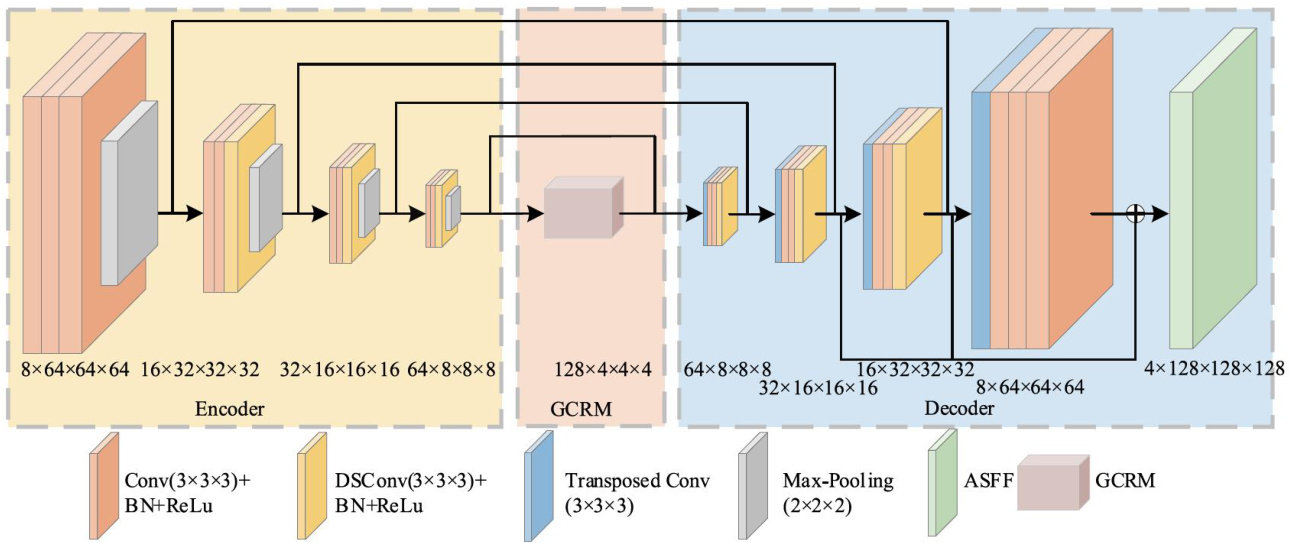
Abstract:Toaddress theisues ofinsuffcient global information capture and inadequatedeep semantic information fusion in the U-Netmodel for MRIbrain tumor segmentation,anovel braintumor segmentation network,ASGU-Net was proposed.
The algorithm wasbasedon 3D U-Net,incorporating agraph convolution inferencemodule tocaptureadditional long-range contextualfeatures.Aditionally,dynamicsnakeconvolution(DSConv)was introduced inthe encoder-decodertobetter accommodate the varied shapes of tumors,enhancing edge feature extractionand improving segmentation acuracy. Furthermore,anadaptivespatial featurefusion(ASFF)modulewas introduced in the decoder toenhance the feature fusion efect byintegrating semantic informationcaptured by multipleencoderblocks.Theevaluationonthepubliclyavailable BraT 2019—2021 datasets shows that the Dice values for whole tumor,tumor core and enhanced tumor are 90.70%/90.70% 1 91.00% , 84.90%/84.00%/88.80% and 77.30%/77.40%/82.50% ,respectively. The experimental results demonstrate the effectiveessofASGU-NetinthebraintumorsegmentationtaskASGU-Netcaneectiveladdressstheissuesofiadequateglobal informationcaptureand feature fusion,providing effective reference for high-precisionautomatedbrain tumorsegmentation.
KeyWords:computer neuralnetwork;brain tumor segmentation;3D U-Net;graph convolution inferencebotteneck layer; dynamic snake convolution;adaptive spatial feature fusion
脑肿瘤是脑部异常细胞形成的肿块,严重威胁人类的健康与生命。(剩余15198字)
