表皮样囊肿破裂的超声图像特征分析

打开文本图片集
Analysis of ultrasound imaging features of ruptured epidermoid cysts
ZHANG Shuangshuang1,CHEN Huyan2,CHEN Shuying³,DING Hong1,LI Xia1,ZHAO Tianyu1,QIAO Xiaohui1 1.Departmentof Ultrasound Medicin,2.Departmentof Dermatology,3.DepartmentofLaboratory,Huashan Hospital, FudanUniversity,Shanghai 2OoO4O,China
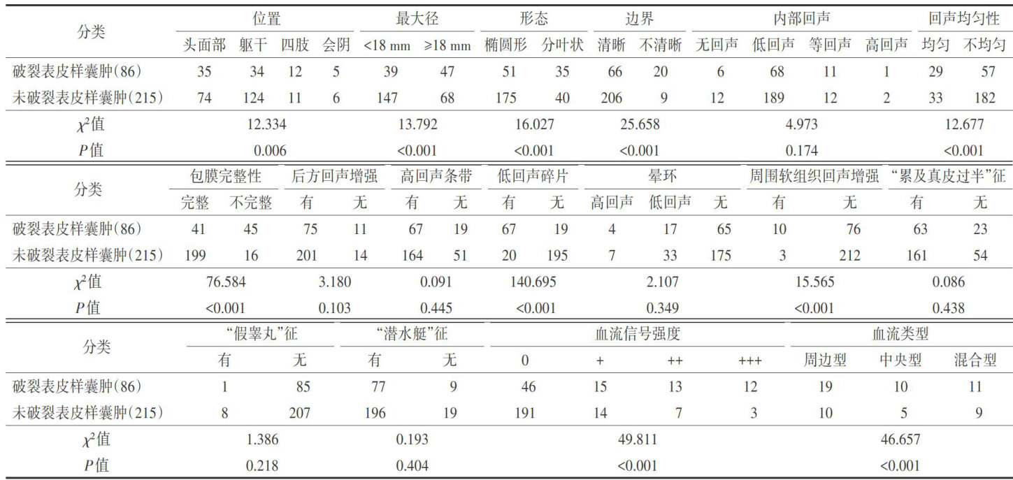
ABSTRACTObjectiveTo analyze and summarize theultrasound imaging features of ruptured epidermoid cysts. MethodsTheultrasoundimagingdataof86rupturedepidermoidcystsand215urupturedepidermoidcystsconfirmedbysurgical pathologywereretrospectivelyanalyzed,ndthediferencesofultrasound imagingfeatures werecompared.ResultsTherewere significantdifrencsinlsonlocatioaiiameer,apeoudarisogeicityomogeityapsuleei presenceoflowchoicebris,urroundngsoftissuechogencityloodflowsignalintensityanddistrbutionpatebewen ruptured and unruptured epidermoid cysts(all P <0.05).Unruptured epidermoid cysts were commonly found in the trunk,with a maximumdiameter<18mm,typicalovalinsape,well-defiedboundaries,ntactcapsules,eterogeneousintealos withoutow-choicdebris,noehancedsurroundingsoft tssueechogenicityandnobloodflowsgnalsobservedinterallyand peripherally.Incontrast,rupturedepidermoidcysts weremorefrequentlylocatedintheheadandneckwithamaximumdiameter> 18mm,lobulatedmorphology,illdefinedboundariesincompletecapsules,homogeneousinteralhosccompanedbylow echoicdebris,andabundantbloodflowsignalsobserved internallyandperipherally,withenhancedsuroundingsofttisue echogenicity.ConclusionRuptured epidermoidcysts exhibit distinctultrasound imaging features.Ultrasoundcan ffectively differentiate ruptured from unruptured epidermoid cysts,with promising clinical value.
KEYWORDSUltrasonography;Epidermoid cysts,ruptured; Imaging features
表皮样囊肿又称表皮囊肿、漏斗部囊肿、表皮包涵囊肿等,是一种常见的皮肤良性肿块1],全身均可发病,常见于头面部、躯干、四肢等部位,少数可发生于男女外生殖器等部位。(剩余5740字)
