不同磁共振成像技术对脑部胶质瘤的诊断分析

打开文本图片集
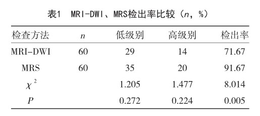
【摘 要】目的 分析不同磁共振成像技术对脑部胶质瘤的诊断效果。方法 选取本院2020年1月-2022年1月收治的60例脑胶质瘤患者为研究对象,均接受磁共振弥散加权成像技术(MRI-DWI)和磁共振波谱成像技术(MRS)检查明确诊断,以手术病理结果为金标准,比较两种检查技术的诊断结果及诊断效能。结果 MRS检出率为91.66%,高于MRI-DWI的71.66%,差异有统计学意义(P
【关键词】磁共振成像技术;脑部胶质瘤;磁共振弥散加权成像
中图分类号:R739.41 文献标识码:A 文章编号:1004-4949(2022)24-0080-03
Diagnostic Analysis of Different Magnetic Resonance Imaging Techniques for Brain Glioma
LI Ze-yuan
(School of Biomedical Engineering and Medical Imaging, Hubei University of Science and Technology, Xianning 437100, Hubei, China)
【Abstract】Objective To analyze the diagnostic effect of different magnetic resonance imaging techniques on brain glioma. Methods A total of 60 patients with glioma admitted to our hospital from January 2020 to January 2022 were selected as the research objects. All patients were diagnosed by magnetic resonance diffusion weighted imaging (MRI-DWI) and magnetic resonance spectroscopy (MRS). The diagnostic results and diagnostic efficacy of the two techniques were compared with the results of surgical pathology as the gold standard. Results The detection rate of MRS was 91.66%, which was higher than 71.66% of MRIDWI, and the difference was statistically significant (P
【Key words】Magnetic resonance imaging technology; Brain glioma; Magnetic resonance diffusion weighted imaging
脑部胶质瘤(brain glioma)根据恶性程度分为低级别、高级别[1]。(剩余4311字)
